
Equipment/Technology update
I wanted to write a blog post on some of the equipment I’ve been building/optimizing in lab, mainly our data acquisition system (DAQ). Our lab uses analog chart recorders to record data from two electrode voltage clamp electrophsiology experiments in Xenopus laevis oocyte because digitizers are expensive (>$5000) but here is a low-cost solution (<$500 including laptop).
Chart recorders
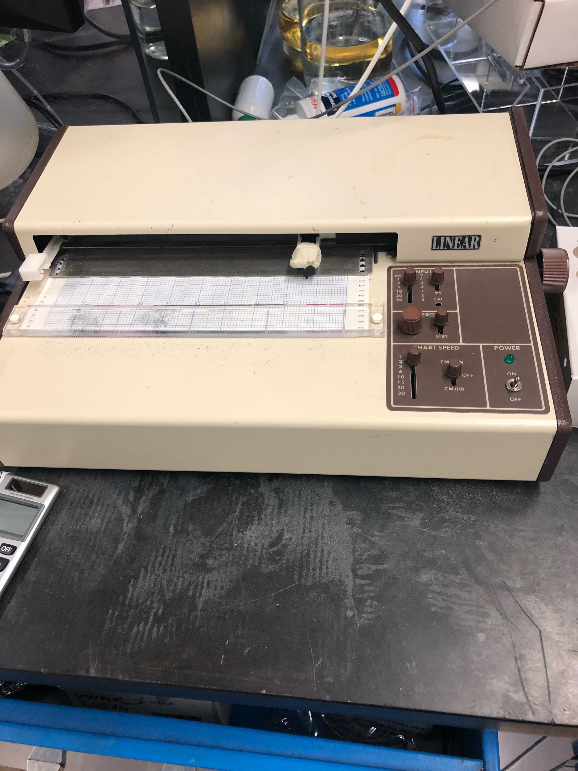
A chart recorder, which “acquires” data from our oocyte clamps/amplifiers using pen and paper.
Up until now, our lab has relied on analog chart recorders to obtain oocyte electrophysiology recordings. These are the kinds of things you would see in old movies whenever a lie detector test appeared on the screen: a moving pen and chart paper. When we record a response (or a change in current) from cells, the oocyte clamp sends the data (“coded” as a change in voltage) out, but from there, its up to the researcher how they acquire (or record) the data. Chart recorders take the voltage from the clamp/amplifier and sends it to a motor that moves an ink pen. This pen “convert” the voltage to movement, which get decoded by the researcher performing the experiment. For example, say an oocyte moves the pen 20 squares. When we have the chart recorder on the 2mV setting, we know that a single square is “worth” 2nA. BUT that’s not the whole story. Our oocyte clamps have an operating range, which we set ahead of time. If our recordings are over or under that range, we can’t get the response. Because we expect really big responses, we scale down the response by a factor (called “gain)” of 10, so when it comes to decoding the data, we have to re-apply this factor int the recordings. Therefore, the 20 squares would be 27*2*10=540nA of current.
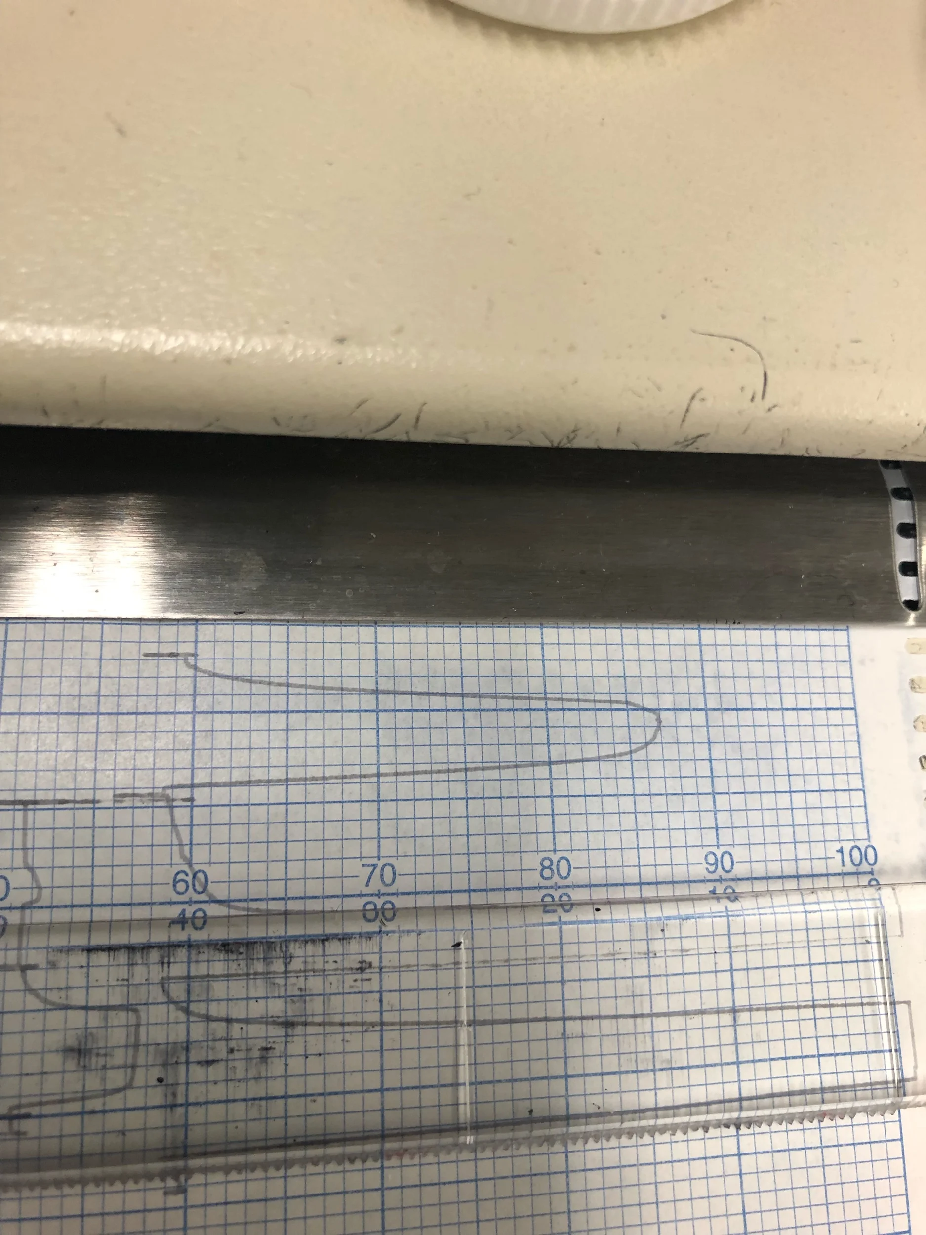
A nice response to 2 uM glutamate. One major drawback of the analog chart recorders is that you have to pick a data range ahead of time, which means if you guess wrong, you’ll either have the pen run off of the chart paper, or get a very tiny tracing.
Digitizers
Analog chart recorders are an inexpensive way to record certain types of data, but it is getting increasingly more difficult to find paper and ink pens to keep them running. For this reason, I set out to find a practical “analog-to-digital converter” (ADC or digitizer.) My personal goal was to keep the system price as low as possible, while making it easy to use, since we have undergraduate and high school students who perform the experiments/recordings. The common solution to this problem is to use molecular devices’ Digidata DAQ systems, although those end up being more expensive than the entire electrophysiology rig. The most cost-effective dedicated oocyte electrophysiology DAQ system I found is about $4,000. On paper, this seems like a better deal, but remember that I’m trying to convert 4 stations, so the real cost of switching systems would be >$16,000, which is still high. Plus, our analog chart recorders still work, so there technically isn’t a whole lot of pressure to update the systems.
When I joined lab, we had two running electrophysiology rigs, and I’ve slowly repaired/built two additional rigs. I’m very proud of the digital microscopes we incorporated, because 1) it makes teaching new students electrophysiology very easy and 2) it reduces eye strain during especially long experiments. My personal mission, however, has always been to update the chart recorders. I always felt that seeing the data on a computer screen, already converted to units of current (amps), would help our students understand what is really going on during experiments. For example, its easy for a student to mistakenly report an experimental result in “squares” which is technically meaningless.
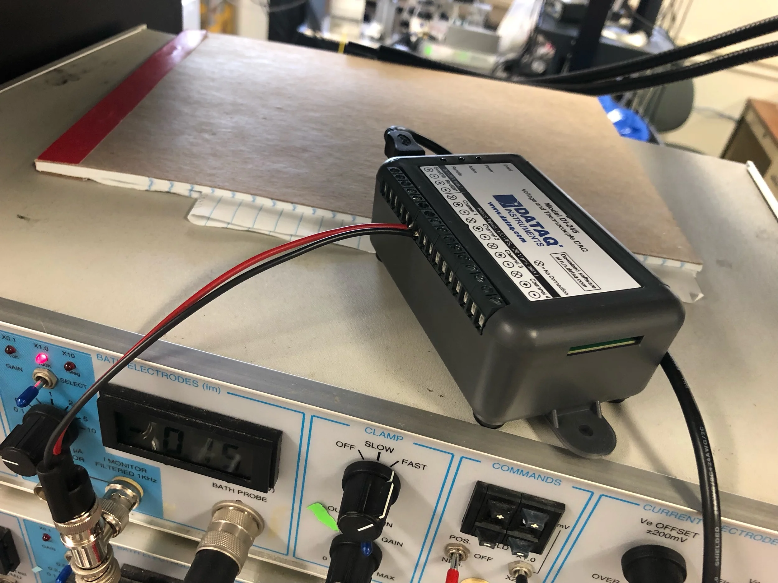
The DATAQ DI-245. Very easy to set up, low noise, and affordable.
To find an alternative, I first looked through the amplifier manual (we use a warner oc725C) and figured out the output range of the system. An important note: we have two sets of gain multiplier switches (0.1, 1, and 10, or 0.1, 0.2, 1, 2, and 10). You can think of the gain setting to scale the output; if you are getting small, hard-to-record responses, multiply the response by a large number or vice versa. These settings have different characteristics, so like everything, you must optimize the settings for what you’re trying to do. The system I decided to go with was the DATAQ instruments DI-245. The hardware itself reads a wide range of voltages and supports a low sample rate (important for oocyte recordings) and the software seemed simple to use. Plus, you can export your data to MS Excel for further analysis. Another selling point: it was only $300. DATAQ has more affordable alternatives too ($60-170), I just wanted a range of values to optimize and work with.
I’m currently in the final stages of optimization/validation, but I can say that it’s more than capable of handling our recordings: it covers our range of currents, and the software lets you mark events, like applying drug or agonist. Best of all, there’s almost no electrical noise!
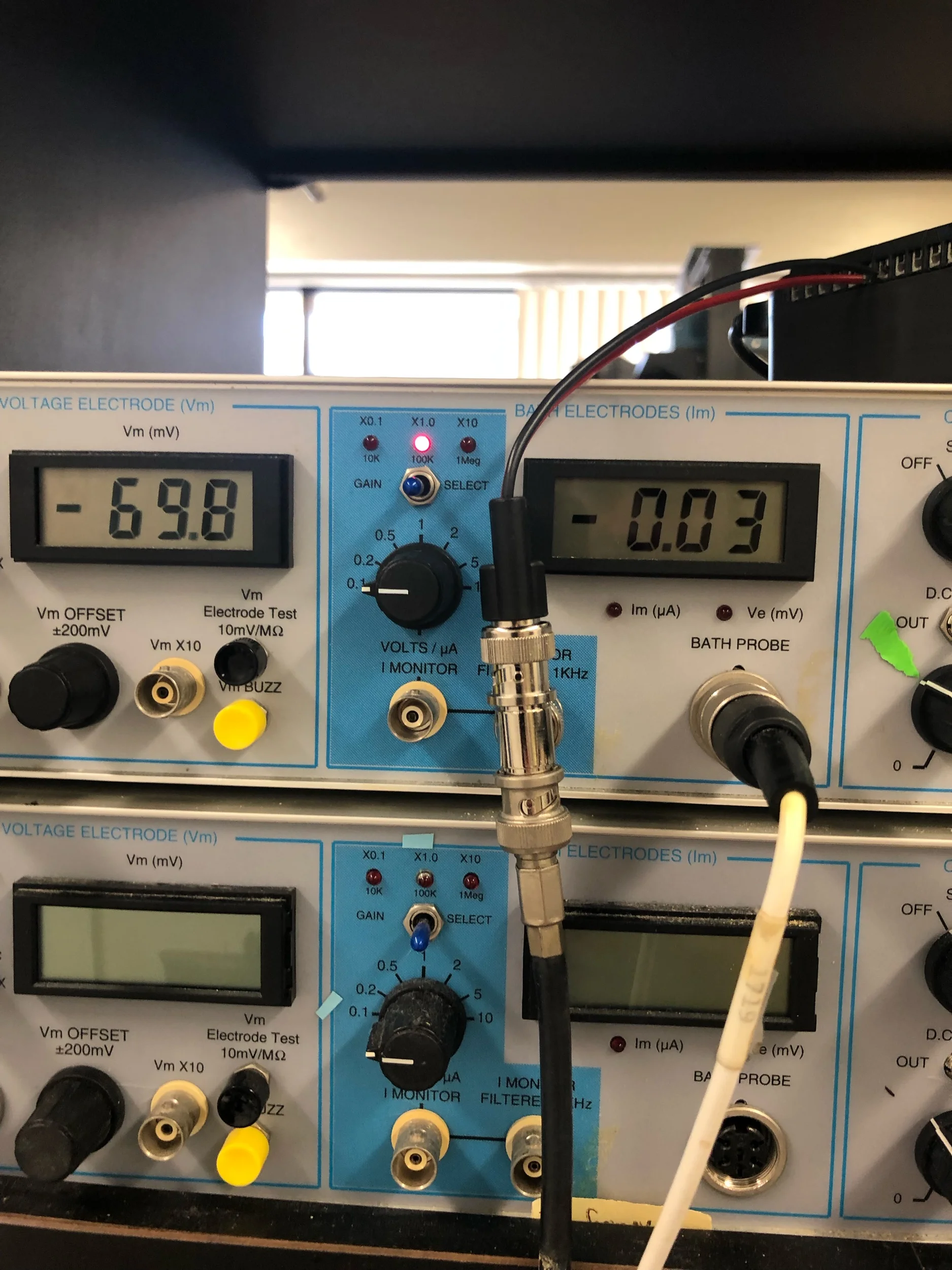
The oocyte amplifier reporting the membrane potential (-70mV) on the left, and the current flowing through the cell to maintain the membrane potential (0.03 uA.)
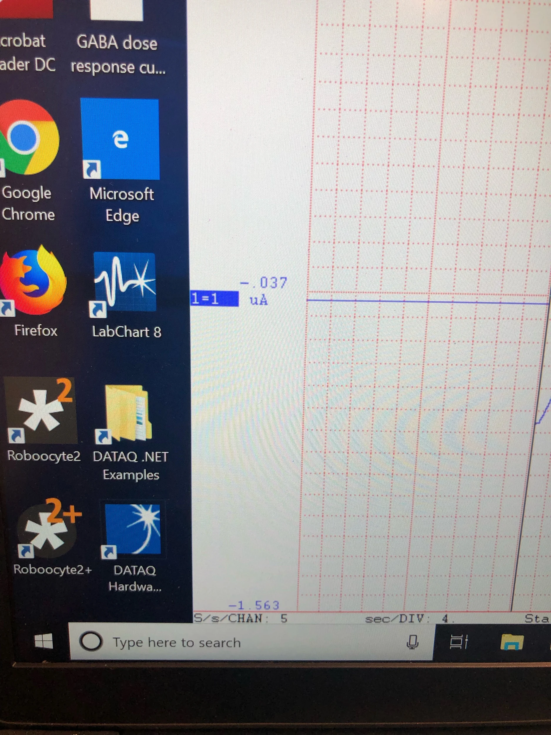
The DATAQ software reporting the current, which matches what the amplifier is reporting (0.03 uA). Before we rely on any new equipment, we have to go through a validation process, meaning that it will produce the same results as what has been in use up to this point.
A previous lab member was having trouble getting a digitizer (an Axon minidigi 1A) to work, even in faraday cage, due to electrical noise in our lab, which interferes with data acquisition. The digitizer was given as a sort of freebie when you purchased the more expensive digidata systems, although using that system today requires the newest software from molecular devices, which is overkill for what we need, and a little on the expensive side.

A very nice tracing recorded using the DI-245. The data from the analog chart recorder and from the DI-245 are essentially identical, and like I mentioned before, essentially no noise!
Future Directions


This originally started off as a side project while my big cloning experiments were in progress, but I’m not done yet. I still want to push our lab into incorporating newer technology, so for me, the next step for me is to try to incorporate raspberry pi into this system. You can build an inexpensive touchscreen interface (<$200) which is cheaper than buying a dedicated laptop. I bought this Elecrow CrowPi pre-assembled system for prototyping and testing, so I’ll keep you posted on how things go. My main concern is memory usage, since the software runs continuously, and any bottlenecks in memory could crash the software, causing us to lose all the data generated in an experiment (a risk in any digital data acquisition system unfortunately.)


